Themen
-
Fachbereich
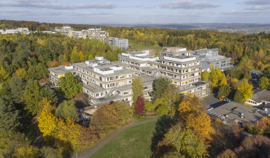
Verwaltung, Organisation und Service am Fachbereich Biologie -

Studium
Ob studieninteressiert oder bereits eingeschrieben - hier finden Sie alle Informationen -

Forschung
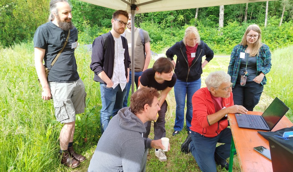
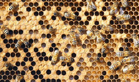
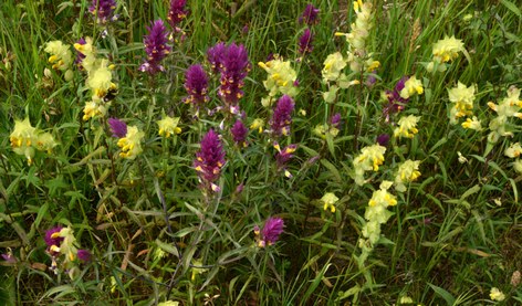
Wir erforschen vielfältige Fragestellungen, die sich mit Molekülen und Zellen über Entwicklung und Funktion bis hin zu Biodiversität und Naturschutz beschäftigen -
Fachgebiete
Die Arbeitsgruppen am Fachbereich sind in 11 Fachgebieten repräsentiert.
-
Fachbereich
Verwaltung, Organisation und Service am Fachbereich Biologie -
Studium
Ob studieninteressiert oder bereits eingeschrieben - hier finden Sie alle Informationen -
Forschung
Wir erforschen vielfältige Fragestellungen, die sich mit Molekülen und Zellen über Entwicklung und Funktion bis hin zu Biodiversität und Naturschutz beschäftigen -
Fachgebiete
Die Arbeitsgruppen am Fachbereich sind in 11 Fachgebieten repräsentiert.