Main Content
CELLS
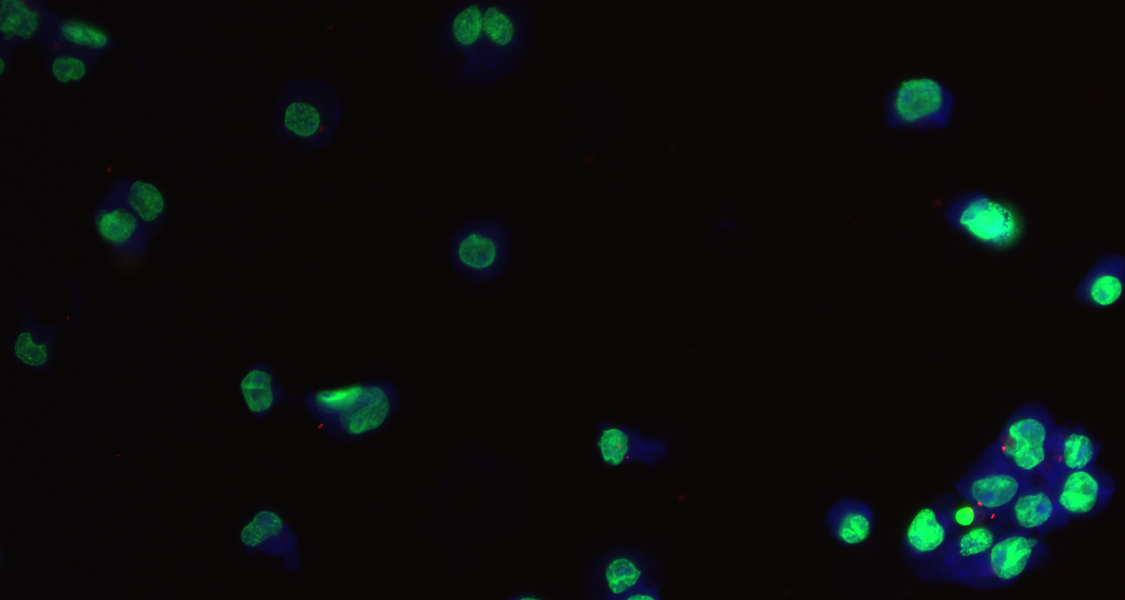
To analyze cell infection and changes in cell morphology in fluorescence microscopy images, proper cell detection and segmentation are required. In automated fluorescence microscopy image analysis, the separation of signals in close proximity is a challenging problem. High cell densities or cluster formations increase the probability of such situations on the cellular level. Another limitation is the detection of morphologically complex cells, such as macrophages or neurons. We provide a challenging dataset of macrophage cells originating from high-throughput screening, together with our deep learning models and the corresponding software. The dataset contains 2552 cells and 2595 nuclei, including ground truth bounding boxes and segmentation masks.