Main Content
The formation of a functional cardiovascular system is essential for the development of all vertebrates. During embryonic development, a network of blood vessels is formed to supply the organs with oxygen, nutrients, signaling molecules, and metabolites, while removing waste products contained in the blood (Time-lapse recording with a spinning disc microscope of a beating heart of a transgenic zebrafish embryo. Red blood cells are genetically labeled with a DsRed (red) and blood vessels with a GFP (green).
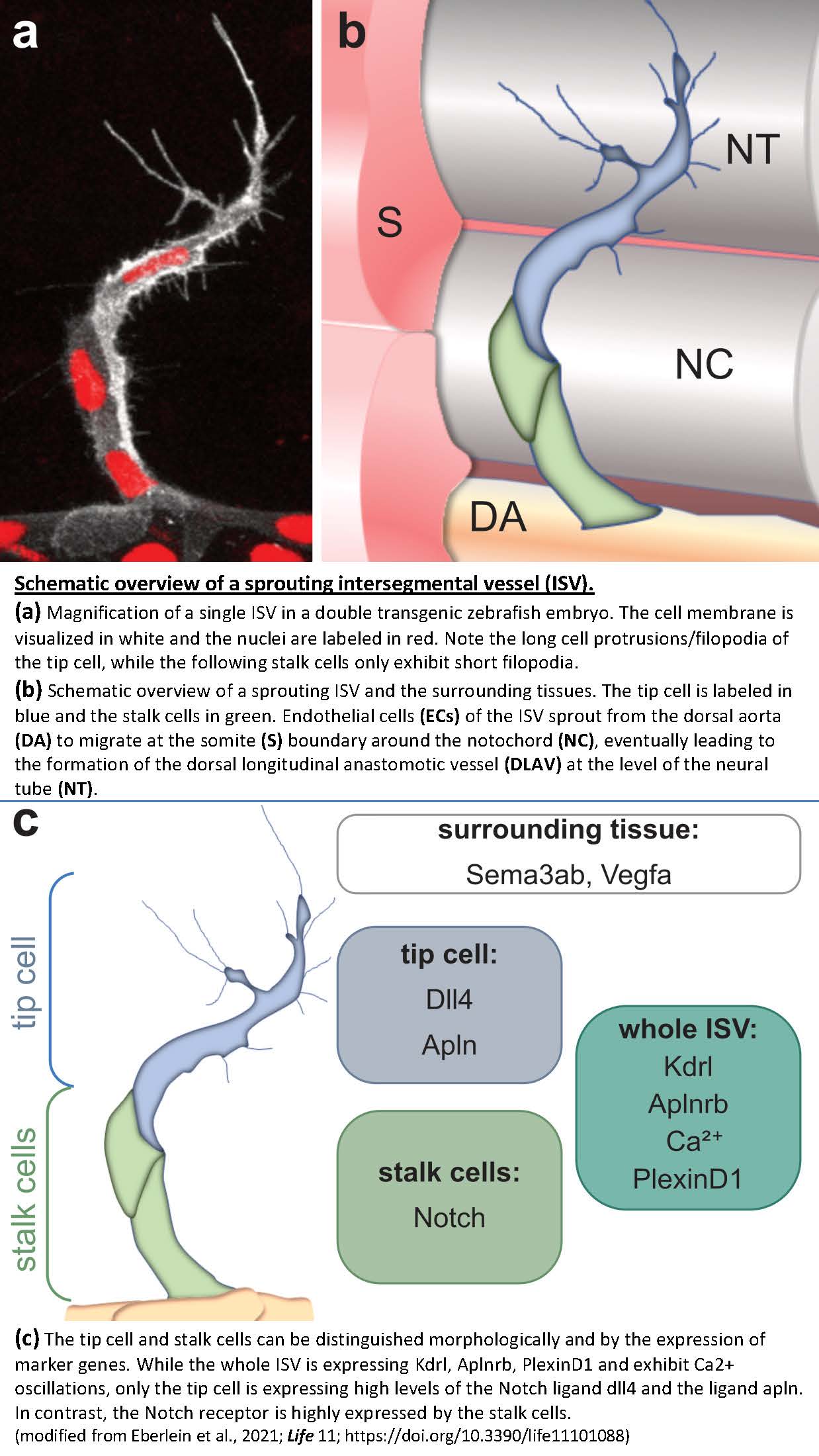
During the development of the cardiovascular system, endothelial cells migrate from existing blood vessels in a process known as angiogenesis and form a network of arteries and veins. In the course of this migration, so-called tip cells and stalk cells form and continuously exchange signals with their environment that control the growth of the new blood vessels. Tip cells are located at the tip of the newly forming vessel and direct the migration of the new blood vessel (Time-lapse recording with a confocal microscope of forming blood vessel of a transgenic zebrafish embryo. Endothelial cells are genetically labeled with a GFP (white) and nuclei of endothelial cells are genetically labeled with an mCherry (red)). Stalk cells, on the other hand, follow the tip cells, proliferate, and form the new lumen. The inner cell layer facing the lumen is formed by endothelial cells surrounded by a basal lamina, pericytes, and smooth muscle cells that regulate vascular tone. The process of angiogenesis not only occurs during embryonic development, but is also reactivated during wound healing/tissue regeneration and in many diseases, especially cancer. In addition to blood vessels, the cardiovascular system also consists of a network of lymphatic vessels. These lymphatic vessels return lymph from the capillary bed to the circulation and play an important role in fluid homeostasis and immunity.