28.10.2026 Mini-Organs and Big Data
How AI Is Advancing Organoid Research
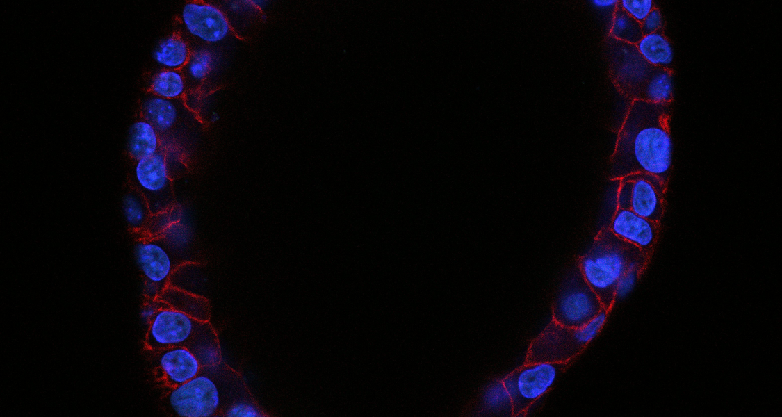
Researchers from Marburg University, Helmholtz Munich, Helmholtz AI and the German Cancer Research Center (DKFZ) aim to create one of the most comprehensive openly accessible image collections of organoids to date. The data will help develop AI models for the automated analysis of these mini-organs, opening up new possibilities for disease research, drug development, and personalized medicine. The German Research Foundation (DFG) is funding the SCOPE-AI project with approximately €330,000 under it’s “Research Data and Software” program.
Organoids and AI: Automating the Analysis of Lab-Grown Mini-Organs
Organoids are three-dimensional miniature models of human and animal organs grown in the lab. They allow researchers to study diseases and test drug candidates, and they can help reduce the need for animal experiments.
Analyzing the resulting microscopy images is still largely done by hand, a process that is laborious, slow, and error-prone. Artificial intelligence could automate this analysis, but large, diverse, and carefully annotated datasets for training powerful models have so far been lacking. This is precisely the gap SCOPE-AI aims to close.
At Marburg University, Prof. Dr. Mareike Lehmann and Prof. Dr. Thomas Worzfeld will contribute to SCOPE-AI by investigating human and mouse lung and kidney organoids, respectively. Their research will focus on applying artificial intelligence to characterize epithelial phenotypes and cellular plasticity in these organoid models. By combining different microscopy techniques with AI-based image analysis, the researchers aim to identify and quantify biologically relevant changes in epithelial cell behavior, differentiation, and organization. A particular focus is the development of AI approaches that enable automated phenotypic characterization. The Marburg contribution will thus help establish innovative, scalable approaches to studying epithelial biology and plasticity across different organs and species.
Open to the Entire Research Community
The project will generate three openly accessible datasets, each tailored to a specific application. All datasets will be released under an open license and hosted on platforms including the NFDI4BIOIMAGE infrastructure and Zenodo. To help researchers use the data right away, the team will also provide baseline AI models, annotation tools, and example workflows. In this way, the project reflects Helmholtz AI’s approach of making AI methods and resources openly and broadly available to the life sciences. “Organoids are a highly promising tool for biomedical research, yet their images are still mostly analyzed by hand. AI can do this faster and more objectively, but it needs high-quality training data, which is exactly what has been missing. With SCOPE-AI, we are making such data openly available across different organs and laboratory conditions. Open, well-documented resources move the entire research community forward,” says Dr. Marie Piraud, Head of the AI Consultant Team for Health at Helmholtz AI, based at Helmholtz Munich, and co-lead of the Clinical AI Consultant Team at AI for Health.
An Interdisciplinary Consortium from Munich, Marburg, and Heidelberg
SCOPE-AI was jointly secured by four principal investigators:
- Dr. Marie Piraud, Helmholtz AI, Helmholtz Munich
- Prof. Dr. Mareike Lehmann, Biomedical Research Center, Philipps-Universität Marburg
- Prof. Dr. Thomas Worzfeld, Institute of Pharmacology, Philipps-Universität Marburg
- Dr. Jens Puschhof, Junior Research Group Epithelium-Microbiome Interactions, German Cancer Research Center (DKFZ), Heidelberg
The consortium combines AI expertise with long-standing experience in organoid, lung, kidney, and intestinal research.
More Information: DFG-GEPRIS: SCOPE-AI