Hauptinhalt
Publikationen von Stephan Imhof
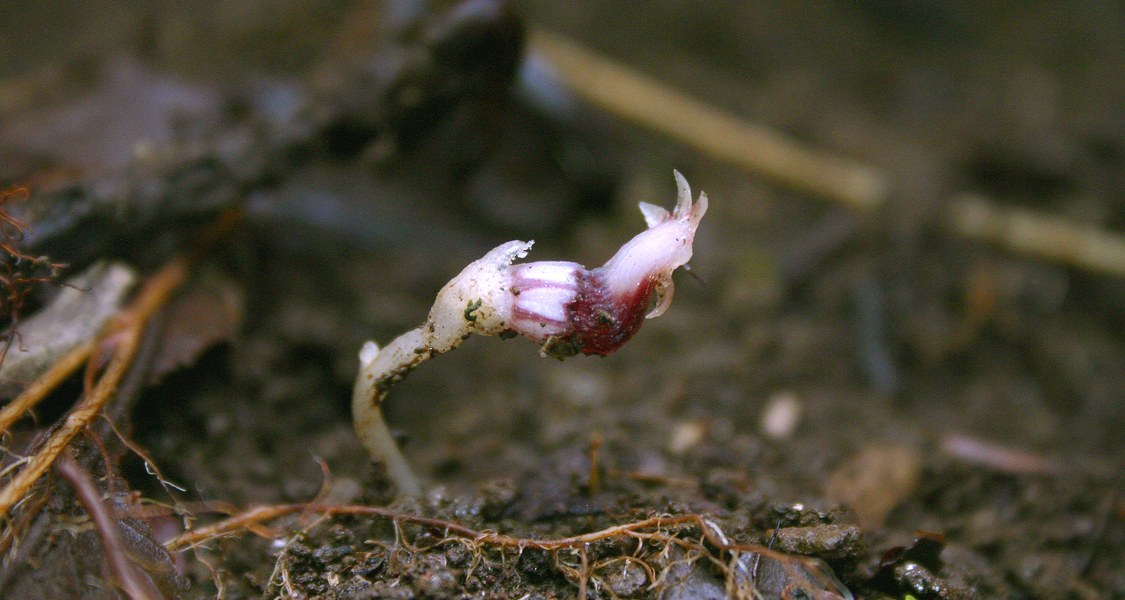
Feller, B., Dančák, M., Hroneš, M., Sochor, M., Suetsugu, K., Imhof, S. (2022): Mycorrhizal structures in mycoheterotrophic Thismia spp. (Thismiaceae): functional and evolutionary interpretations. Mycorrhiza online
Achlorophyllous, mycoheterotrophic plants often have an elaborate mycorrhizal colonization pattern, allowing a sustained benefit from external fungal root penetrations. The present study reveals the root anatomy and mycorrhizal pattern of eight mycoheterotrophic Thismia spp. (Thismiaceae), all of which show separate tissue compartments segregating different hyphal shapes of the mycorrhizal colonization, as there are intact straight, coiled and peculiarly knotted hyphae as well as degenerated clumps of hyphal material. Those tissue compartments in Thismia roots potentially comprise exo-, meso- and endoepidermae, and exo-, meso- and endocortices, although not all species develop all these root layers. Differences in details among species according to anatomy (number of root layers, cell sizes and shapes) and colonization pattern (hyphal shapes within cells) are striking and can be discussed as an evolutionary series towards increasing mycorrhizal complexity which roughly parallels the recently established phylogeny of Thismia. We suggest functional explanations for why the distinct elements of the associations can contribute to the mycorrhizal advantage for the plants and, thus, we emphasize the relevance of structural traits for mycorrhizae. (open access)
Imhof, S., Feller, B., Heser, A. (2020): Morpho-anatomical differences among mycoheterotrophic Afrothismia spp. (Thismiaceae) indicate an evolutionary progression towards improved mycorrhizal benefit. Mycorrhiza 30(2): 397-405
Achlorophyllous, mycoheterotrophic plants depend on their mycorrhizal fungi for 100% of their carbon supply. Hence, there is strong evolutionary pressure towards a well-organized functioning of the association from the plant’s perspective. Members of the mycoheterotrophic genus Afrothismia have evolved elaborate fungal colonization patterns allowing a sustained benefit from external fungal penetration events. On the basis of anatomical details of the root-shoot systems of A. korupensis and A. hydra, we elucidate an evolutionary progression between the comparatively simple mycorrhizal pattern in A. gesnerioides and the so far most complex mycorrhiza in A. saingei. We detected two major advancements: (1) two species, A. korupensis and A. saingei, use the fungus itself as energy storage, replacing starch depositions used by A. gesnerioides and A. hydra, and (2) the morphological complexity of hyphal forms in plant tissue compartments increases from A. gesnerioides to A. saingei. We discuss the omitting of starch metabolism as well as the morpho-anatomical differences as an evolutionary fine-tuning of the compartmented mycorrhizal organization in Afrothismia. Our results support the idea of a taxonomic distinction between Afrothismia and other Thismiaceae. (Open Access)Imhof, S. (2017): Distel, Wein und Pfaffenhütchen - Die Pflanzendarstellungen am Deckengewölbe der Kugelkirche. Pinguindruck, Berlin.
Zur 500-Jahr-Feier der Kugelkirche in Marburg wurde ich gebeten, die Deckenornamente botanisch einzuschätzen. Aus diesen Betrachtungen ist ein kleines Heft mit Bestimmungsversuchen geworden. Dieses ist im Eingangsbereiche der Kugelkirche für wenig Geld zu erwerben.
Rath, M., Grolig, F., Haueisen, J., Imhof, S. (2014): Combining microtomy and confocal laser scanning microscopy for structural analysis of plant-fungus associations. Mycorrhiza 24(4): 293-300
The serious problem of extended tissue thickness in the analysis of plant–fungus associations was overcome using a new method that combines physical and optical sectioning of the resin-embedded sample by microtomy and confocal microscopy. Improved tissue infiltration of the fungal-specific, high molecular weight fluorescent probe wheat germ agglutinin conjugated to Alexa Fluor® 633 resulted in high fungus-specific fluorescence even in deeper tissue sections. If autofluorescence was insufficient, additional counterstaining with Calcofluor White M2R or propidium iodide was applied in order to visualise the host plant tissues. Alternatively, the non-specific fluorochrome acid fuchsine was used for rapid staining of both, the plant and the fungal cells. The intricate spatial arrangements of the plant and fungal cells were preserved by immobilization in the hydrophilic resin Unicryl™. Microtomy was used to section the resin-embedded roots or leaves until the desired plane was reached. The data sets generated by confocal laser scanning microscopy of the remaining resin stubs allowed the precise spatial reconstruction of complex structures in the plant–fungus associations of interest. This approach was successfully tested on tissues from ectomycorrhiza (Betula pendula), arbuscular mycorrhiza (Galium aparine; Polygala paniculata, Polygala rupestris), ericoid mycorrhiza (Calluna vulgaris), orchid mycorrhiza (Limodorum abortivum, Serapias parviflora) and on one leaf–fungus association (Zymoseptoria tritici on Triticum aestivum). The method provides an efficient visualisation protocol applicable with a wide range of plant–fungus symbioses. (https://doi.org/10.1007/s00572-013-0530-y)
Imhof, S., Massicotte, H.B., Melville, L.H., Peterson, R.L. (2013): Subterranean morphology and mycorrhizal structures. In: Merckx, V.S.F.T. (ed.): Mycoheterotrophy - The Biology of Plants Living on Fungi. Springer Verlag, Heidelberg, pp. 157-215
In autotrophic plants, many scientific questions can be dealt with using generalized concepts of root structure and function (e.g. Kutschera and Lichtenegger 1992; Polomski and Kuhn 1998; Gregory 2006). However, this certainly does not hold for mycoheterotrophic (MH) plants. The structure of roots, rhizomes, or subterranean scale leaves of MH plants intimately linked to the association with soil fungi is of critical ecological relevance because these plants essentially depend upon fungi for their carbon and perhaps other nutrient needs. Hence, the subterranean organs of MH plants often show remarkable morphological and anatomical adaptations to meet their speci fi c requirements. This chapter, therefore, addresses the importance of morphology and anatomy to complement modern methods for understanding the fungal colonization patterns in MH plants and their relationships to function.
Rath, M., Weber, H.C., Imhof, S. (2013): Morpho-anatomical and molecular characterization of the mycorrhizas of european Polygala species. Plant Biology 15: 548-557
The mycorrhizas of 12 species of Polygala (Polygalaceae), including herbs, subshrubs and one shrub, collected from Germany, Mallorca (Spain) and Malta, were investigated by morpho‐anatomical and molecular methods. Aseptate hyphae, arbuscules and vesicles indicate an arbuscular mycorrhiza in all species examined. Hyphal spread in Polygala is predominantly, but not exclusively, intracellular and comprises three characteristic stages of colonization: (i) intracellular, linear hyphal growth in a cascading manner after penetration towards the penultimate parenchyma layer (layer 2), (ii) initially linear hyphal growth in the cells of layer 2 from where hyphal branches repeatedly penetrate the anatomically distinct innermost parenchyma layer (layer 1), forming arbuscule‐like structures therein which are subject to degeneration, (iii) more branches from the linear hyphae in layer 2 develop, but coil and make contact to the layer outside layer 2 (layer 3) in which arbuscule‐like structures similar to those in layer 1 form and degenerate. This general colonization pattern differs in details between the species, and critical comparisons, in particular between the woody P. myrtifolia, the herbaceous Polygala spp. and the mycoheterotrophic Epirixanthes spp. (Polygalaceae) suggest an evolutionary shift of mycorrhizal features within the family towards an optimization of plant benefit through the fungus. Based on the molecular marker 18S rDNA mycorrhizal fungi detected in roots of Polygala spp. are largely restricted to five clades of Glomeraceae 1 (Glomus Group A). This result rejects the hypothesis of a strict symbiotic specificity in Polygalaceae but may stimulate a discussion on functionally compatible groups of fungi. (https://doi.org/10.1111/j.1438-8677.2012.00680.x)
Imhof, S. (2010 onwards): Mycoheterotrophic plants - How many of them are there? Scratchpads - Biodiversity online
Scratchpads is a content management system that allows to assemble taxonomic data (and more) of a given field and present it to interested readers. I have gathered the accepted names and their synonyms of non-orchid mycoheterotrophic plants, bibliographic data of about 1400 publication from 1753 until today associated with mycoheterotrophy, and provide short paragraphs on taxonomic history and diagnoses of the species. Data collection on mycoheterotrophic orchids are in progress. Learn more under http://mhp.myspecies.info
Imhof, S. (2010): Are monocots particularly suited to develop mycoheterotrophy? In: Seberg, O., Petersen, G., Barfod, A., Davis, J. I. (eds): Diversity, Phylogeny and Evolution in the Monocotyledons. Aarhus University Press, Kopenhagen, pp. 11-23
More than 400 plant species are mycoheterotrophic, of which by far the majority are monocotyledons (88%) – a remarkable statistic, considering monocots account for only about 23% of all flowering plant species. Which factors make monocotyledons so appropriate for this mode of life? Subterranean organs of several non-related mycoheterotrophic plants share common features, which in turn can be interpreted as either supporting, or even being prerequisites for, the mycoheterotrophic habit. These are: (1) herbaceous habit, (2) a maintained and often voluminous primary root cortex parenchyma, (3) a tendency for a star-like organisation of the root system, mostly with short and thick or long and thin roots, and (4) an often complicated mycorrhizal colonization pattern. Monocots meet these putative demands for mycoheterotrophy better than other angiosperms for the following reasons: (1) Monocots are primarily herbaceous. (2) Monocot roots lack secondary growth, which normally obliterates the primary cortex in dicots. (3) The secondary homorhizy of monocots predetermines a starlike root system, which in dicots must be achieved by reductions of allorhizy. (4) Monocots are able to protect their central cylinder through a comparatively inexpensive tertiary endodermis. (5) Due to their predisposition for shoot-borne roots and multilayered cortices, monocots are more prone to develop voluminous and diverse subterranean organs (rhizomes, scale leaves, roots), which in turn promote the development of complex colonization patterns. Feel free to ask for a copy.
Imhof, S. (2009): Arbuscular, ecto-related, orchid mycorrhizas - three independent structural lineages towards mycoheterotrophy. Implications for classification? Mycorriza 19: 357-363.
The classification of mycorrhizas in seven equally ranked types glosses over differences and similarities and, in particular, does not acknowledge the structural diversity of arbuscular mycorrhizas. This article emphasizes the parallel continua of ecto-related mycorrhizas and arbuscular mycorrhizas, exemplified within Ericaceae and Gentianales, respectively, as well as the proprietary development of orchid mycorrhizas, all three of which have independently developed mycoheterotrophic plants. A hierarchical classification according to structural similarities is suggested. (https://doi.org/10.1007/s00572-009-0240-7)
Appelhans, M., Weber, H.C., Imhof, S. (2008): Rutaceae sampled from Germany, Malta and Mallorca (Spain) are associated with AMF clustering with Glomus hoi (Berch & Trappe). Mycorrhiza 18: 263-268
Six Rutaceae species collected from natural habitats (Malta, Mallorca (Spain), and Tenerife (Spain)) and the Botanical Garden in Marburg were examined with respect to mycorrhizal structures and fungal identity. All species have the same gross colonization pattern of arbuscular mycorrhiza (AM) with distinct intracellular and intercellular phases but show remarkable differences in details, especially in terms of the extent of the intracellular phase. The associated AM fungi, identified using molecular methods, cluster together with Glomus hoi Berch & Trappe, although the plants were collected from very distant locations. (https://doi.org/10.1007/s00572-008-0179-0)
Imhof, S., Sainge, M.N. (2008): Ontogeny of the mycoheterotrophic Afrothismia hydra (Burmanniaceae). Bot. J. Linn. Soc. 157:31-36
The complete ontogeny of the mycoheterotrophic Afrothismia hydra (Burmanniaceae) from seed to seed dispersal is presented. The, oblong-ovoidal seeds are up to 0.7 mm long. They germinate with root tissue only, disrupting the seed coat and developing a primary ovoid root tubercle. At the proximal end of the tubercle a second tubercle arises, and further root initials indicate the sequential growth of more root tubercles with filiform extensions resulting in a small root aggregate. The seed coat often remains attached to this structure. When the root aggregate enlarges a central axis to which all roots are connected becomes visible. This axis has a growth pole where new root tubercles arise. The same growth pole will later develop into a stem with scale leaves finally terminating in a flower. Flowers develop sympodially when the mature plant is only several centimetres long. After anthesis, the corolla tube disintegrates, leaving a pyxidium which opens by means of an peculiar elongating placenta, here called ‘placentophore’. The placentophore elevates the placenta with attached seeds above the flowering level and is interpreted as an adaptation to ombrohydrochory. The reduction of hypocotyl, cotyledon and primary shoot is discussed with regard to the classical germination concepts of monocotyledons , and with mycoheterotrophic dicotyledons. (https://doi.org/10.1111/j.1095-8339.2008.00787.x)
Imhof, S. (2007): Specialized mycorrhizal colonization pattern in achlorophyllous Epirixanthes spp. (Polygalaceae). Plant Biol. 9: 786-792
Roots of the achlorophyllous Epirixanthes papunana and E. elongata were sectioned in complete series in order to reconstruct the three-dimensional mycorrhizal colonization pattern within their tissues. Hyphal morphology, vesicles, as well as the exclusively intracellular mode of colonization indicate a Paris-type of arbuscular mycorrhiza showing a hitherto unknown colonization pattern: (1) the outer cortex is colonized by persistent straight-growing hyphae which branch in a cascading manner, (2) a specific layer (called layer 2) is inhabited by persistent hyphal coils, (3) in the cells of the anatomically distinct inner cortex parenchyma layer (called layer 1) the hyphae immediately degenerate, and (4) the layer outside to layer 2 (called layer 3) is either transitional layer 2 when penetrated from the outer cortex or the fungal material degenerates when colonized from the layer 2. This complex colonization pattern is a reasonable adaptation to the particular demands of Epirixanthes as a myco-heterotrophic plant. It not only allows a sustained benefit from the fungal symbiont but also provides a two-level distribution system of hyphae within the roots. The outer cortex hyphae function as a permanent intraradical resource of living fungi providing connection to the external mycelium as well as a coarse distribution of hyphae within the root. Layer 2 represents the fine scale distribution of hyphae, having access to all potentially digesting cells of the layers 1 and 3. Common structural features of mycorrhizae in myco-heterotrophic plants are pointed out in order to find putative prerequisites for their heterotrophic mode of life. (https://doi.org/10.1055/s-2007-965613)
Imhof, S. (2006): Two distinct fungi colonize roots and rhizomes of the myco-heterotrophic Afrothismia gesnerioides (Burmanniaceae). Can. J. Bot. 84: 852-861
The subterranean organs of Afrothismia gesnerioides H. Maas consist of short rhizomes densely covered with ovoid root tubercles, each of which may extend into a short filiform root extension. Serial sections revealed the presence of two distinct fungi occupying different niches within the plant tissues. Rhizomes and roots are divided into separate compartments hosting different morphotypes of the aseptate, exclusively intracellular hyphae of fungus A: (i) straight and persistent hyphae in the root epidermis, root extension, and outer rhizome cortex, (ii) coiled but still persistent hyphae in the third root layer, (iii) coiled hyphae undergoing degeneration in the root cortical parenchyma, (iv) starch depositions in the inner rhizome cortex and no colonization by fungus A, and (v) a partly collapsed root hypodermis serving as compartment barrier. The colonization by fungus A is interpreted as an aberrant arbuscular mycorrhiza of the Paris type. The compartmentation allows the separation of tissues where the hyphae stay functional from those in which the fungal material is digested. This pattern may represent a complex but efficient strategy for a sustained benefit from the few fungal penetrations that occur. Comparison with earlier work on Afrothismia winkleri (Engl.) Schltr. revealed considerable differences between the mycorrhizae that are interpreted as evolutionary steps. There are signs that these changes even may have improved the mycorrhizal benefit for the plant. The monomorphic hyphae of fungus B are smaller in diameter, septate, grow inter- as well as intra-cellularly, but are always characteristically appressed to the inner cell walls. It does not change its appearance within the root/rhizome compartments as does fungus A. Neither hyphal degeneration nor interferences with fungus A, starch depositions, or alterations in the development of A. gesnerioides could be noticed. Fungus B possibly is a commensal, but relevance to the symbiosis cannot be ruled out. (https://doi.org/10.1139/b06-024)
Imhof, S. (2005): Fundmeldungen 1196.-1200. Botanik und Naturschutz in Hessen 18: 75-77
Hinweise auf Vorkommen in Hessen und angrenzenden Gebieten von Atriplex micrantha, Cerinthe minor, Galeopsis pubescens, Genista pilosa, Gypsophila muralis.
Imhof, S. (2004): Fundmeldungen 1128.-1133, Buchbesprechung, Website-Empfehlungen. Botanik und Naturschutz in Hessen 17: 138-140, 173-174, 179-183
Hinweise auf Vorkommen in Hessen von Bupleurum rotundifolium, Euphorbia maculata, Hydrocotyle vulgaris, Lathyrus hirsutus, Scandix pecten-veneris, Setaria verticillata, Valerianella rimosa. Buchbesprechung von Lüder, R. (2004): Grundkurs Pflanzenbestimmung. Quelle & Meyer, Wiesbaden. Von den Website-Empfehlungen ist heute nur noch die Angiosperm Phylogeny Website online und nach wie vor hoch geschätzt.
Imhof, S. (2004): Morphology and development of the subterranean organs of the achlorophyllous Sciaphila polygyna (Triuridaceae). Bot. J. Linn. Soc. 146: 295-301
The subterranean organs of the achlorophyllous Sciaphila polygyna (Triuridaceae) are described, depicted, and structurally explained for the first time. Unlike other Triuridaceae, the subterranean system of S. polygyna appears as a complex star-like structure of short but thickened roots as well as scale leaves and shoots. A complete series of sections revealed the following construction: in the axil of a scale leaf at a shoot of first order a side shoot of second order as well as a pair of endogenous shoot borne-roots arise. This side shoot of second order also develops a scale leaf very early in ontogeny, which again gives rise to a side shoot of third order and a pair of shoot-borne roots. Other scale leaves at shoots of any order may also bear shoots and root pairs. This growth pattern occurs in a very close manner without internode elongation, resulting in the clumped, star-like appearance. The described structures superficially resemble the root systems of many mycoheterotrophic plants from other families. Comparisons with respect to how they develop, however, show that these similar root systems can result from distinct developmental patterns, suggesting independent evolutionary pathways and a considerable evolutionary pressure towards abbreviated and thickened roots in mycoheterotrophic plants. Possible advantages as well as evolutionary implications of the described structures are discussed. (https://doi.org/10.1111/j.1095-8339.2004.00333.x)
Wu, W.Y., Weber, H.C., Imhof, S. (2004): Morphogenesis and structure of hairs of Tetrapanax papyriferus (Hook.) K. Koch (Araliaceae). Beitr. Biol. Pflanzen 73: 213-221
The green parts of Tetrapanax papyriferus (Hook.) Koch (Araliaceae) are covered by complex, morning star like, multicellular trichomes. These trichomes initially appear as papilla-like cells of the epidermis, which develop into multicellular, sometimes biseriate stalks. At maturity the stalks are crowned by a star-like structure, consisting of many cells with ray-like protrusions in a globose arrangement, the protrusions radiating into all directions. Altready in early stages of the hairs, these heads are present as morula-like cell agglomerations. The central part of the gead eventually appears brownish, caused by secretory activity.
The hair cover is most prominent on both sides of young leaves, the abaxial sides of older leaves and bracts, on peduncle, pedicels, the upper third of the abaxial side of petals as well as the hypanthium of the inferior ovary. The hair cover on the adaxial surfaces of the older leaves, bracts and petals as well as the upper part of the ovary around the styles is less dense to absent. At the upper part of the ovary the hairs are additionally characterized by having shorter stalks but more rays.
The irritant indumentum, covering the susceptible young and reproductive organs, particularly the lower sides of the leaf organs protecting the buds, is discussed as an adaptation primarily against herbivores. (PDF available)Imhof, S. (2003): A dorsiventral mycorrhizal root in the achlorophyllous Sciaphila polygyna (Triuridaceae). Mycorrhiza 13: 327 - 332
The star-like root system of the achlorophyllous Sciaphila polygyna (Triuridaceae) consists of roots up to 1.4 mm thick and 1 cm long seemingly radiating from a single origin. Internally, the roots show a bilateral symmetry when viewed in cross-section: the third root cell layer contains rather loose coils of the aseptate mycorrhizal fungus from the dorsal to the lateral sides, in contrast to the extremely dense coils of thin hyphae in its ventral part. Additionally, the hyphae develop vesicle-like swellings mainly in the central part of the dorsal side as well as the lateral parts of the third layer. The fourth root layer is anatomically heteromorphic, having exceptionally large cells, reaching up to 320×130 m in size (giant cells), in the lower lateral parts. The root-colonizing hyphae only degenerate in the fourth layer, most readily in the giant cells, where they may swell to 24 m in diameter, collapse and end as amorphous clumps. Hyphae in the third layer keep their definite structure.
The structures are interpreted to be the result of a dynamic reaction of the root to the actual fungal penetration points in order to maximize the benefit from the subsequent colonization by compartmentation of the root tissue. The function of the third layer is to host the fungus and keep it alive within its cells, while mainly the giant cells serve for its digestion.
Many indications suggest an arbuscular mycorrhiza for this association. Similarities and differences to other myco-heterotrophic species are discussed. (https://doi.org/10.1007/s00572-003-0255-4)Döring, M., Imhof, S., Weber, H.C., Ewald, D. (2003): Propagation by leaf cuttings of Eleutherococcus Max. (Araliaceae). J. Appl. Bot. 77: 57-60
Eight species of the important medicinal and ornamental genus Eleutherococcus (Araliaccae) were tested upon their ability to regenerate roots and shoots from detached mature leaves in a minimal nutrition Solution. Additionally, leaflets, leaflets with their petiolules detached, and leaves with abscised leaf bases of E. gracilistylus were also tested, accordingly.
The capability for adventitious root development at leaves differed between species. Leaf cuttings of E. divaricatus, E. gracilistylus, E. seoulensis, E. koreanus and E. sieboldianus regenerated abundant adventitious roots at their petioles and survived up to 8 months (E. gracilistylus). From E. lasiogyne only one sample grew minute roots , but died off little later than the other samples of that species. E. henryi did develop callus tissue, but failed to grow adventitious roots and wilted soon. E. lasiogyne x E. sessiliflorus failed to regenerate roots as well as callus tissue at all. Leaflets with or without petiolules as well as leaves with detached bases of E. gracilistylus developed adventitious roots, just like the regular leaf cuttings of this species. Under white light none of all samples grew an adventious shoot, although the leaf cuttings which develop adventitious roots may stay alive for many months. However, leaves of E. gracilislylus, treated the same as all other samples but kept under red light, did develop adventitious shoots: first time in Araliaceae. Since according to the literature most other woody plants also failed to propagate from leaf cuttings so far, our results should be of interest for growers and breeders looking for a cheap and preserving proliferation method.Imhof, S. (2001): Subterranean structures and mycotrophy of the achlorophyllous Dictyostega orobanchoides (Hook.) Miers (Burmanniaceae). Rev. Biol. Trop. 49(1): 237 - 245
Plants of Dictyostega orobanchoides arise from about 1 mm thick rhizomes, which are densely covered by sessile, imbricate, peltate scale leaves. The resulting interfoliar spaces are inhabited by fungal hyphae up to 6 µm thick, often developing vesicle-like bladders. The fungus also colonizes the tissue of the scale leaves, inter- as well as intracellularly, forming vesicles but no arbuscules, and it even penetrates the vascular bundles of the leaves. The rhizome itself does not become infected. The 200 µm thick roots emerge from the rhizome and have a 2-layered cortex and voluminous rhizodermis, which both are delicate and often disrupted or missing. In contrast, the strongly reinforced, tertiary endodermis and the central cylinder are durable and have a considerable tensile strength. Although the roots grow through the hyphal masses in the interfoliar spaces when emerging from the rhizome, they only become infected from the rhizosphere. A collar of rhizomogenous tissue hinders the interfoliar hyphae from direct contact to the roots. Only within the rhizodermis, the mycorrhizal fungus builds coils of heteromorphic hyphae, arbuscule-like structures, and vesicles. Hence, the mycorrhiza in D. orobanchoides is assigned to the arbuscular mycorrhiza. It is hypothezised, that the ephemeral mycorrhizal tissue combined with the durable vascular system of the roots is a strategy to avoid the high costs of protecting the large rhizodermal surface area. The rhizomogenous collar is explained as an extra protection to the tender, young roots, when emerging from the rhizome. The necessity to include other subterranean plant organs along with the roots in future mycorrhizal studies is emphasized. (PDF available)
Diallo, A., Weber, H.C., Imhof, S. (2001): Sonderstellung der Mykorrhiza von Mercurialis perennis L. im Vergleich mit anderen Euphorbiaceae. Beitr. Biol. Pflanzen 72: 315-323
Mercurialis perennis shows a Paris-type VAM, characterized through an exclusively intracellular colonization of the cortex cells by coiled hyphae. Arbuscules are mostly developed laterally as reduced small shrub-like structures at the hyphal coils. Vesicles were not found. In contrast, Euphorbia pulcherrima Willdenow, Manihot esculenta Crantz, Pedilanthus tithymaloides L. and Ricinus communis L. show an Arum-type VAM, with a predominantly intercellular spread of the hyphae, developing large intracellular arbuscules as lateral branches and intracellular as well as intercellular vesicles. Due to its extratropical distribution as well as many morphological features, Mercurialis perennis is to be considered as an advanced member of its family. The finding of a Paris-Type in Mercurialis perennis and Arum-Typs VAM in the other Euphorbiaceae therefore support the previously suggested hypothesis of a phylogenetically more advanced position of the Paris-Type VAM.
Imhof, S., Weber, H.C. (2000): Root structures and mycorrhiza of the achlorophyllous Voyria obconica Progel (Gentianaceae). Symbiosis 29: 201 - 211
The genus Voyria comprises 19 achlorophyllous, mycotrophic species with reduced cormi. Roots of Voyria obconica are up to 1 cm long, 1 - 1.5 mm thick, succulent, brittle and radiated from the shoot base, forming a star-shaped root system. In cross section the central cylinder consists of up to 10 central vessels, surrounded by some parenchymatous cells, 5 to 7 strands of phloem and a pericycle. The cell walls of the anatomically inconspicuous endodermis are characterised by a faint suberin lamella. The cortex is divided into an inner cortex, with 3 to 5 layers of longitudinally elongated cells and a multilayered outer cortex, comprising isodiametric cells. The 2-3 cell layers of the dermal tissue also show a faint suberin lamella within their thickened cell walls. Non-parasitic, achlorophyllous plants need symbiotic interactions with mycorrhizal fungi. In V. obconica the exclusively intracellular hyphae of a single mycorrhizal fungus grows after penetration of the dermal tissue straight towards the inner cortex. Within the inner cortex the hyphae proceed parallel to the central cylinder. Branches of these straight inner cortex hyphae then colonize the outer cortex, where they form coils, swell, and eventually degenerate to amorphous clumps. Similarities and differences in root structure and mycorrhiza to the closely related Voyria tenella are elucidated. Arguments are given to call this association a special form of a Paris-type arbuscular mycorrhiza. The ecological significance of the revealed mycorrhizal compartmentation is discussed. (PDF available)
Imhof, S. (1999): Anatomy and mycotrophy of the achlorophyllous Afrothismia winkleri (Engl.) Schltr. (Burmanniaceae). New Phytol. 144: 533-540
Afrothismia winkleri develops fleshy rhizomes, densely covered with small root tubercles, narrowing to filiform roots with age. The exclusively intracellular mycorrhizal fungus has distinct morphologies in different tissues of the plant. In the filiform root the hyphae grow straight and vesicles are borne on short hyphal stalks. The straight hyphae are present in the epidermis of the root tubercles, but change to loosely coiled and swollen hyphae in the rhizome tissue. No penetration from epidermis to root cortex was found. From the rhizome, a separating cell layer permits only one or rarely two hyphal penetrations into the cortex of each root tubercle. The hyphae proceed apically within the root hypodermis in a spiral row of distinctively coiled hyphae, branches of which colonize the inner root cortex. In the inner root cortex the hyphal coils degenerate to amorphous clumps. In older roots the cortex itself also deteriorates, but epidermis, hypodermis, endodermis and central cylinder persist. The mycorrhizal pattern in A. winkleri is interpreted as an elaborate exploitation system whereby the fungus provides carbon and nutrients to the plant and, simultaneously but spatially distinct, its hyphae are used to translocate and store the matter within the plant. Several features indicate that the endophyte is an arbuscular mycorrhizal fungus. (https://doi.org/10.1046/j.1469-8137.1999.00532.x)
Imhof, S. (1999): Subterranean structures and mycorrhiza of the achlorophyllous Burmannia tenella Bentham (Burmanniaceae). Can. J. Bot. 77: 637-643
Plants of the myco-heterotrophic Burmannia tenella Benth. form star-shaped root systems consisting of 0.7 - 2 mm thick, succulent, brittle roots, reaching lengths of up to 3 cm. In cross section the roots consist of an epidermis, about 10 layers of parenchymatous cortex cells, an endodermis with u-shaped secondary cell wall depositions and a very reduced central cylinder with two to five central xylem elements and two opposite phloem strands, surrounded by a pericycle of relatively large cells.
Based on the thick, aseptate, intracellularly coiled hyphae, arbuscules, and the frequent vesicles, the fungal association of B. tenella is considered to be a Paris-type arbuscular mycorrhiza. The morphological and anatomical structures of the root are discussed in the context of the mycorrhizal dependency of B. tenella.
In some root samples, a second fungus with septate hyphae colonized the cortex intracellularly. This fungus restricts the spread of the aseptate symbiont without causing morphological changes to the cortex cells. (https://doi.org/10.1139/b99-034)Imhof, S. (1999): Root morphology, anatomy and mycotrophy of the achlorophyllous Voyria aphylla (Jacq.) Pers. (Gentianaceae). Mycorrhiza 9: 33-39
Roots of V. aphylla only develop hairs where roots of neighbouring plants or organic litter is attached. Fungal penetrations almost exclusively occur at these root-to-root attachments. The ecological significance of these immediate hyphal bridges for achlorophyllous plants is discussed. The morphological and anatomical features of the root of Voyria aphylla, as well as its Paris-type arbuscular mycorrhiza (AM) appear to be transitional between those of V. truncata and V. tenella. A hypothetical evolutionary progression of AM and its significance for the development of myco-heterotrophy is proposed. (https://doi.org/10.1007/s005720050260)
Imhof, S. (1998): Subterranean structures and mycotrophy of the achlorophyllous Triuris hyalina (Triuridaceae). Can. J. Bot. 76 : 2010-2019
Triuris hyalina, an unusual achlorphyllous plant, was investigated for subterranean morphology, root anatomy and mycotrophy. Stems with scale leafs extend subterraneously to a depth of 15 cm. Pairs of adventitious roots develop at the scale leafs and clumps of apparently radiating roots, formed by accumulations of side shoot and scale leaf developments, occur. Roots consist of epidermis, short cell exodermis, three distinct layers of cortex parenchyma, endodermis and an extremely reduced central cylinder with one or two central tracheidal xylem elements.
The fungus associated with Triuris hyalina roots, exhibit thick walled, 6-9 µm thick, aseptate external hyphae. It penetrates the epidermis by developing appressoria and enters the cortex solely through the short cells of the exodermis. In the cortex cells, the aseptate hyphae start to coil. In the outer cortex layer hyphae are thin, frequently branched and most densely coiled. In the middle cortex layer they are thicker, less densely coiled and mostly appear degenerated to clumps of amorphous fungal material. The inner cortex layer rarely becomes colonized. Vesicles occur in the outer and the middle cortex layers. This mycorrhizal pattern is interpreted as an adaption to attain a sustainable use from the endophyte. It is suggested that the mycorrhiza in Triuris hyalina be interpreted as a type of arbuscular mycorrhiza (AM). Implications for systematics and ecology are discussed. (https://doi.org/10.1139/b98-168)Weber, H.C., Alexander, S., Imhof, S. (1998): The enrichment of ginseng garden soils with vesicular-arbuscular mycorrhizal fungi. In: Weber, H.C., Zeuske, D., Imhof, S.: Ginseng in Europe. Proceedings of the 1st European Ginseng Congress, pp 59-66. Marburg, Germany, 6-11 December 1998
Panax species are highly dependent on vesicular-arbuscular mycorrhiza (VAM). However, due to their mycorrhizal form (called Paris-type VAM), ginseng species decrease the vigor of their endophytes and jeopardize the fungal support for the long term. The introduction of additional VAM propagules is therefore necessary. This paper suggests the use of a VAM fungus wet nurse plant in ginseng gardens in order to maintain the mycorrhizal potential of the soil at a high level. The wet nurse plant should meet the following requirements: the plant roots should be highly susceptible for mycorrhizal fungi, the mycorrhiza should be the Arum-type VAM, and, the plant should be readily accessible and inexpensive to maintain.
Our investigations reveal, that for the mid-latitudes of the northern hemisphere, all requirements are met by Epilobium angustifolium (Onagraceae) and its introduction into ginseng gardens is recommended.Imhof, S. (1997): Root anatomy and mycotrophy of the achlorophyllous Voyria tenella Hooker (Gentianaceae). Bot. Acta 110: 298-305
The central cylinder of the root of Voyria tenella consists of up to ten central, non-lignified, tracheidal xylem elements surrounded by some parenchymatic tissue and 5-7 groups of phloem. A pericycle could not be discerned. Even though the endodermis carries a faint suberin lamella it can not be discerned anatomically without special staining. The cells of the 1-3 cortex layers next to the endodermis are elongated longitudinally, the subsequent cortex parenchyma is multilayered and consists of isodiametric cells. The cells of the 2-3 layered outer dermal tissue are smaller than those of the adjacent cortex, their walls carry a suberin lamella and the outermost of them constantly scale off. The dermal tissue is interpreted as a multilayered exodermis.
The fungal colonization in roots of Voyria tenella remarkably differs from any known mycorrhizal pattern. After having penetrated the dermal tissue, the always intracellularly growing hyphae head straight towards the inner cortex layers, where they spread along the central cylinder. Ramifications from these inner spreading hyphae then colonize the cortex parenchyma from the inside and they develop dense hyphal coils. Eventually, the coiled hyphae swell and collapse, resulting in amorph clumps of fungal material. This mycorrhizal pattern is referred to as an intraradical fungus garden.
Arguments are given to call the mycorrhiza in Voyria tenella a specialized arbuscular mycorrhiza. Phylogenetic and ecological implications of the observations and the results are discussed. (https://doi.org/10.1111/j.1438-8677.1997.tb00643.x)Imhof, S., Weber. H.C. (1997): Root anatomy and mycotrophy (AM) of the achlorophyllous Voyria truncata (Standley) Standley & Steyermark (Gentianaceae). Bot. Acta 110: 127-134
Roots of Voyria truncata retain the primary root structure, even though they can grow as thick as 2 mm in diameter. These root diameters are due to a retained capability for cell division in the cortex parenchyma. This is explained as a vital adaptation to its life form.
Based on the extraradical mycelium, the mode of penetration, the structurally incompatible intraradical phase, the presence of intercellular vesicles in the root cortex, and the occurance of immediate hyphal bridges from arbuscular mycorrhizal roots of neighboring plants, the mycorrhiza of V. truncata is described as an arbuscular mycorrhiza (AM), although the characteristic arbuscles are missing. Special features of the AM in V. truncata are interpreted as an improved efficiency in taking advantage of the mycorrhiza.
Root connections with roots of neighbouring plants are common and preferred locations for fungal infections. An evolutionary tendency towards parasitism of higher plants is discussed. (https://doi.org/10.1111/j.1438-8677.1997.tb00619.x)Imhof, S., Weber, H. C., Gómez, L.D. (1994): Ein Beitrag zur Biologie von Voyria tenella Hook. und Voyria truncata (Standley) Standley & Steyermark (Gentianaceae). Beitr. Biol. Pflanzen 68: 113-123
The chlorophyll lacking Gentianaceae Voyria tenella Hook. and Voyria truncata (Standley) Standley & Steyermark have been observed within their natural habitat, and new results concerning ontogeny and ecology were revealed.V. tenella is annual. The earliest stage of development found, is a structure of only 2 mm in length and apparently constitutes root material. Proceeding from this stage, a morning star type of root-system is developed. Subsequently, 3 to 4 stems are brought out successively. Within a week, the primarily nodding buds come to flower; anthesis lasts only for some days. After 2 months approximately, the life cycle of an individual is accomplished.
In contrast, the roots of V. truncata are relatively strong, numerous and grow more or less horizontale down to 20 cm under the soil surface. Lateral roots up to the 3rd order could be ascertained. Stems are exclusively root-borne and can be branched subterraneously already. Rhizomes do not occur.
Both species are probably ant-dispersed. The developmental abbreviations of the subterranean organs are discussed with respect to phylogenetic tendencies. (PDF available)