Main Content
The chorioallantoic membrane (CAM) assay for the study of head & neck vascular anomalies
Vascular anomalies (VA) are a clinically and histopathologically heterogeneous group of benign lesions. They include hemangiomas, as well as vascular malformations such as lymphatic, venous and arteriovenous malformations (1). Patients with lymphatic and arteriovenous malformations in particular are frequently not only cosmetically-stigmatized, but also functionally-compromised (2). Depending on the anatomical site of the lesion, patients can develop problems with speech, swallowing and breathing, the latter often requiring a tracheostomy (3). Therapy options are limited and do not always achieve functionally- and cosmetically-satisfying results (4, 5). New therapies are thus needed in order to more effectively treat these lesions. In contrast to malignant tumors, however, there are only few experimental systems available for the study of VA. Here, we evaluated the chorioallantoic membrane (CAM) assay, a well-established ex vivo model in cancer and angiogenesis research (6, 7), regarding its suitability as a model system for VA (Figures 1 and 2).
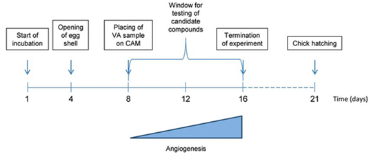
Figure 1. Time scheme, depicting the procedure and highlighting the time window of vessel growth which can be exploited for testing pharmaceutical candidate compounds. (8)
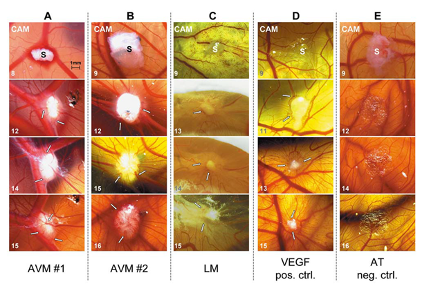
Figure 2. Vascular anomalies (VAs) induce angiogenesis in the chorioallantoic membrane (CAM) assay. Representative micrographs of the three tested VA specimens are depicted in A-C. All arteriovenous malformation (AVM) tissues (A, B), as well as the lymphatic malformation (LM) specimen (C), induced angiogenesis (arrows). A sponge soaked with vascular endothelial growth factor (VEGF) served as a positive control (D), whereas adipose tissue (AT) (E) did not induce any angiogenic reaction. S, Specimen. Numbers in the lower left corner of each image correspond to the respective days of egg development. A scale bar is shown for size comparison. (8)
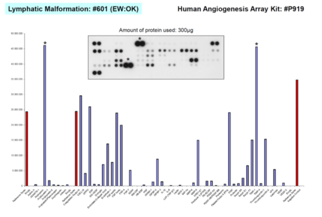
Figure 3. Identification of candidate genes using the Human Angiogenesis Array.
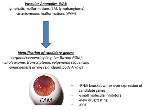
Figure 4. Use of the CAM assay for therapeutic testing. Identified candidate genes (e.g. in Figure 3) can either be inhibited or overexpressed in VA tissues and evaluated in the CAM assay.
In summary, our observations indicate the CAM assay to be a suitable model system for the study of VAs, as well as to show how treatment with pro- and antiangiogenic drugs affects VA growth patterns (Figure 4). The CAM assay has the potential to become a valuable tool for VA studies. The current participants in the CAM project are depicted in Figure 5.
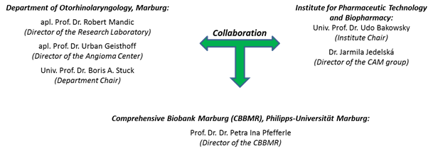
Figure 5. Current participants collaborating within the framework of the CAM project.
Literature
1 Mulliken JB and Glowacki J: Hemangiomas and vascular malformations in infants and children: A classification based on endothelial characteristics. Plast Reconstr Surg 69: 412-422, 1982.
2 Drepper H: Clinical aspects and therapy of lymphangiomas, hemangiomas and nevi in the area of the head and neck. HNO 33: 293-302, 1985 (in German).
3 Buckmiller LM, Richter GT and Suen JY: Diagnosis and management of hemangiomas and vascular malformations of the head and neck. Oral Dis 16: 405-418, 2010.
4 Marler JJ and Mulliken JB: Current management of hemangiomas and vascular malformations. Clin Plast Surg 32: 99-116, ix, 2005.
5 Eivazi B, Ardelean M, Baumler W, Berlien HP, Cremer H, Elluru R, Koltai P, Olofsson J, Richter G, Schick B and Werner JA Update on hemangiomas and vascular malformations of the head and neck. Eur Arch Otorhinolaryngol 266: 187-197, 2009.
6 Hagedorn M, Javerzat S, Gilges D, Meyre A, de LB, Eichmann A and Bikfalvi A: Accessing key steps of human tumor progression in vivo by using an avian embryo model. Proc Natl Acad Sci USA 102: 1643-1648, 2005
7 Vogel HB and Berry RG: Chorioallantoic membrane heterotransplantation of human brain tumors. Int J Cancer 15: 401-408, 1975.
8 Jedelská J, Strehlow B, Bakowsky U, Aigner A, Höbel S, Bette M, Roessler M, Franke N, Teymoortash A, Werner JA, Eivazi B, Mandic R. The chorioallantoic membrane assay is a promising ex vivo model system for the study of vascular anomalies. In Vivo. 2013 Nov-Dec;27(6):701-5.