Main Content
Experimental Methods and Simulations
On this page, we provide an overview of the research methods used in our group. A detailed list of all research instruments and their specifications can be found on the pages of the core facilities of mar.quest.
Growth
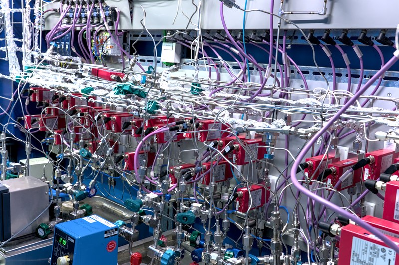
For the growth of novel quantum materials, we use metal-organic chemical vapor deposition (MOCVD) reactors. Precursor gases are introduced into the reactor chamber, where the ratios of the precursor gases and the temperature of the chamber can be precisely controlled so that the precursors decompose in a targeted manner and the desired quantum material grows. The goal is to achieve growth that is as homogeneous as possible over large areas. In addition, we investigate novel precursor gas mixtures with regard to their suitability.
Characterization
In our group, we characterize both the materials we grow ourselves and samples provided by our collaboration partners.
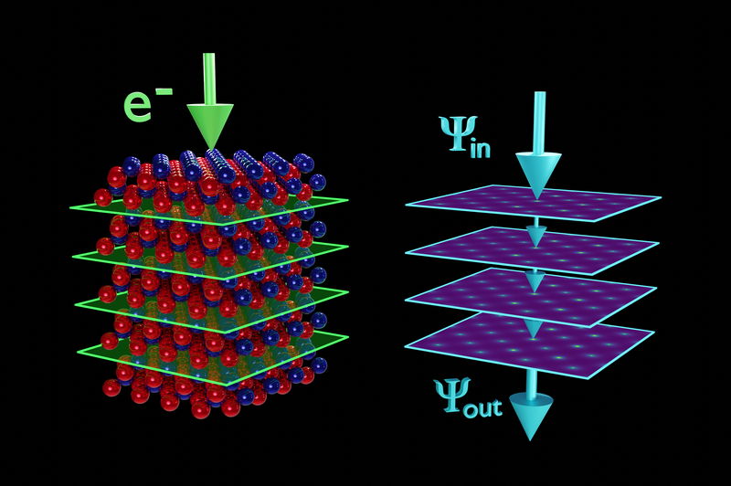
Transmission Electron Microscopy
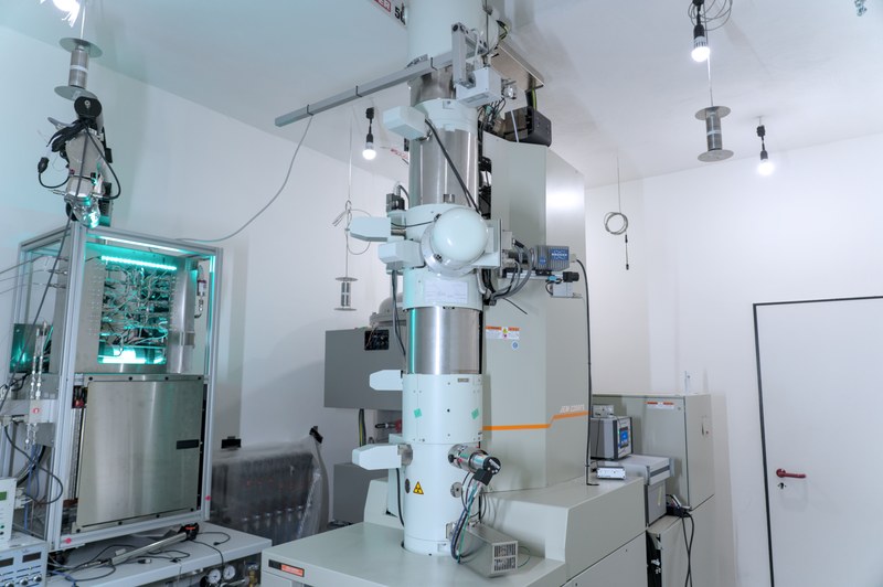
The central characterization method of our research group is (scanning) transmission electron microscopy. We operate two high-performance transmission electron microscopes, and an additional, highly advanced microscope is currently being installed. With our microscopes, it is possible to resolve the atomic structure of our samples in imaging mode. Measurements in diffraction mode provide information on the orientation and phase of the samples. Furthermore, spectroscopic techniques such as energy-dispersive X-ray spectroscopy and electron energy-loss spectroscopy yield insights into the chemical composition of the sample as well as the nature of the chemical bonding.
Focused Ion Beam Preparation and Electron Microscopy
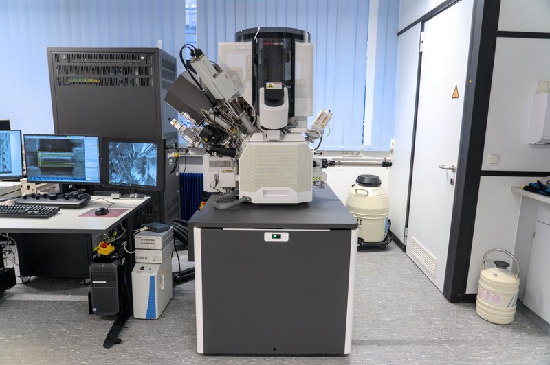
For the preparation of thin samples to be examined with transmission electron microscopes, we use, among other techniques, focused ion beam (FIB) milling. Our dual-beam systems are equipped with both an ion column and an electron column. The electron beam allows investigation of the samples down to the nanometer scale, while the ion beam is used to manipulate the sample. By introducing precursor gases, sub-micrometer-precise protective or connecting layers can be grown to secure the lamellae—only a few micrometers in size and thinned by the ion beam—onto the sample holders for transmission electron microscopy.
Further Methods and Machines
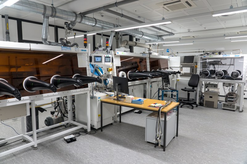
In addition to our electron microscopes, we have access to many other instruments and measurement techniques. Our samples are stored in nitrogen- or argon-filled glove boxes, which prevent degradation from exposure to moisture and air. Using airtight transfer capsules or microscope holders, we can move samples between the glove boxes and the microscopes without contact with the ambient air. Furthermore, we employ techniques such as Raman spectroscopy and atomic force microscopy to study the phase, crystallinity, and morphology of our grown quantum materials. Photoluminescence spectroscopy provides information about the electronic structure of the materials we have grown.
Simulations und Use of AI
To gain a better understanding of our measurement data and to compare experiment with theory, we perform scanning transmission electron microscopy simulations. We also use DFT calculations to compute the absorption spectra of our samples and compare them with experimentally obtained spectra. This allows us to draw conclusions about the crystal phases actually present in our samples. In addition, we employ artificial intelligence to analyze diffraction patterns, enabling us to determine the orientation and crystal phase of the samples.