Main Content
Lymphatic filariasis
In collaboration with the Institute for Medical Microbiology, Immunology and Parasitology (IMMIP) of the University Hospital Bonn and the Kwame Nkrumah University of Science and Technology (KNUST) in Kumasi, Ghana, the aim of our research is to elucidate the genetic causes of lymphatic filariasis.
Clinical and genetic features of lymphatic filariasis
In developing countries, around 120 million people are infected with filaria or roundworms, which can lead to lymphatic filariasis. The WHO ranks this disorder as being among the "most neglected diseases". The majority of infected individuals develop either no, or at most mild, symptoms with recurrent fever. However, some infected individuals display a serious clinical presentation, characterized by the key symptom of lymphedema (LE), or a hydrocele (see Figure 1).
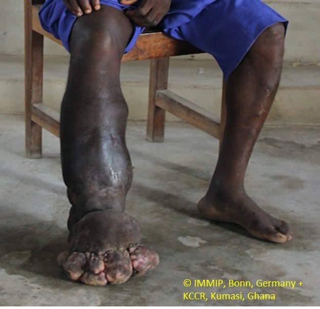
Figure 1: Roundworm larvae enter the human body via a mosquito bite, and develop into adult worms that settle in the lymphatic vessels. In general, the worms are located in the lymphatic vessels of the legs. However, they may also occur in the chest, arms, or genitals. This colonization disrupts normal lymphatic drainage. Over a period of several years, the affected body parts begin to swell. The worms also trigger continuous inflammatory reactions, and inflict substantial damage on the lymphatic system. In cases of chronic infection, LE occurs, whereby the swelling no longer subsides at all. When the lower extremity is affected (a), this manifests as so-called elephantiasis. This term refers to the enormously enlarged leg circumference. If the testicular area is affected, male patients present with inflammation and swelling of the testicles, epididymis, and spermatic cord. This leads to an accumulation of serous fluid in the testicles (hydrocele testis) and lymphedema of the scrotum (lymphatic scrotum). Image provided courtesy of the Institute of Medical Microbiology, Immunology, and Parasitology (IMMIP) of the University Hospital of Bonn.
Around 7% of infected individuals develop LE, and around 30-50% of infected men develop a hydrocele testis (including Tisch et al. (2001) J Infect Dis). Both LE and a hydrocele testis are negatively correlated with the number of filariae in the body. Researchers therefore suspect that both disorders occur as part of the immune defense against the parasite, and are an expression of inflammation (including Pfarr et al. (2009) Parasite Immunol). Research suggests that the genetic variability of the host is of etiological importance. Formal genetic studies have been unanimous in demonstrating a familial tendency for infection with filarias, the number of filariae in the body, and the development of an LE or a hydrocele testis (including Cuenco et al. (2009) J Infect Dis). Lymphatic filariasis is a multifactorial disease, whose etiology involves a large number of genetic risk factors.
To identify the first genetic risk factors for lymphatic filariasis, we are currently conducting a genome-wide association study (GWAS) of a large case-control collective from Ghana. The study is expected to generate deeper insights into the pathophysiology of this serious disorder.
Contact persons
Prof. Dr. Johannes Schumacher
Selected Publications
Buerfent BC, Gölz L, Hofmann A, Rühl H, Stamminger W, Fricker N, Hess T, Oldenburg J, Nöthen MM, Schumacher J, Hübner MP, Hoerauf A. Transcriptome-wide analysis of filarial extract-primed human monocytes reveal changes in LPS-induced PTX3 expression levels. Sci Rep. 2019 Feb 22;9 (1):2562.