07.04.2021 Redox Enzymes of the Thioredoxin Family as Potential and Novel Markers in Pemphigus
New publication of PEGASUS research group
Redox Enzymes of the Thioredoxin Family as Potential and Novel Markers in Pemphigus
P. Sliwiak, E. Folwarczny , D. Didona , S. Fink, C. Wiegand , E. M. Hanschmann , M. Hertl and C. Hudemann
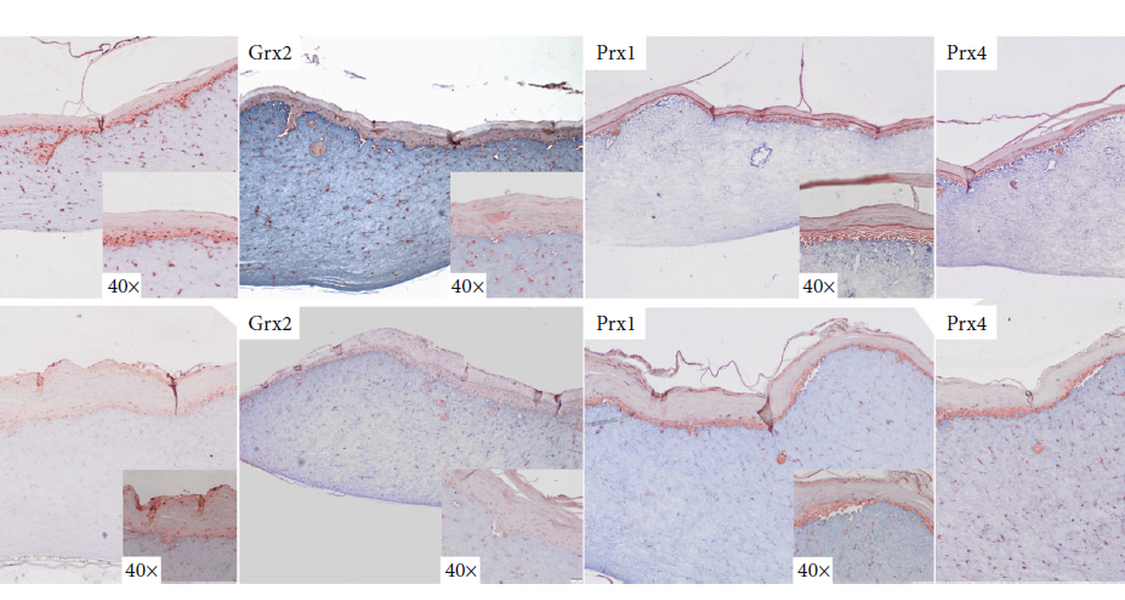
Pemphigus vulgaris (PV) is a severe autoimmune blistering disease affecting both skin and mucous membranes. Its pathogenesis is related to IgG autoantibodies primarily targeting the cellular adhesion protein desmoglein (Dsg) 3, one of the major desmosome components. Impaired redox regulation is considered a major player in the pathogenesis of autoimmune diseases such as pemphigus by enhancing inflammation and breakdown of immunological tolerance by structural protein modifications. Despite many recent advances, local and systemic redox profiles that characterize the immune response in pemphigus are virtually unknown but potentially crucial in further advancing our understanding of redox-dependent modifications that eventually lead to clinical manifestation. Here, we have analyzed the individual expression pattern of four major redox enzymes that are members of the thioredoxin (Trx) fold superfamily (peroxiredoxins (Prxs) 1 and 4, glutaredoxin (Grx) 2, and Trx1) in serum and PBMCs as well as their distribution in the skin of pemphigus patients compared to healthy controls. We show that in groups of five pemphigus patients, Prx1 is upregulated in both serum and PBMCs, while its epithelial distribution remains within the spinous epithelial layer. Expression of Grx2 and Prx4 is both reduced in serum and PBMCs, while their distinct and similar expression in the skin changes from an even distribution throughout the basal layer (healthy) to ubiquitous nuclear localization in pemphigus patients. In PV patients, Trx1 is secreted into serum, and cellular distribution appears membrane-bound and cytosolic compared to healthy controls. We furthermore showed that a 3D ex vivo human skin model can indeed be used to reproduce similar changes in the protein levels and distribution of redox enzymes by application of cold atmospheric plasma. Deciphering the relationship between redox enzyme expression and autoimmunity in the context of pemphigus could be critical in elucidating key pathogenic mechanisms and developing novel interventions for clinical management.
For the original publication Click here
Contact
Christoph Hudemann, PhD
Tel.: +49 (0) 6421 - 58 - 64823
Mail: hudemanc@staff.uni-marburg.de
Philipps University Marburg
Department of Dermatology
Baldingerstraße
D-35043 Marburg, Germany