Main Content
TP6new: Desmosomal hyper-adhesion and heterophilic desmoglein interactions in pemphigus
Summary
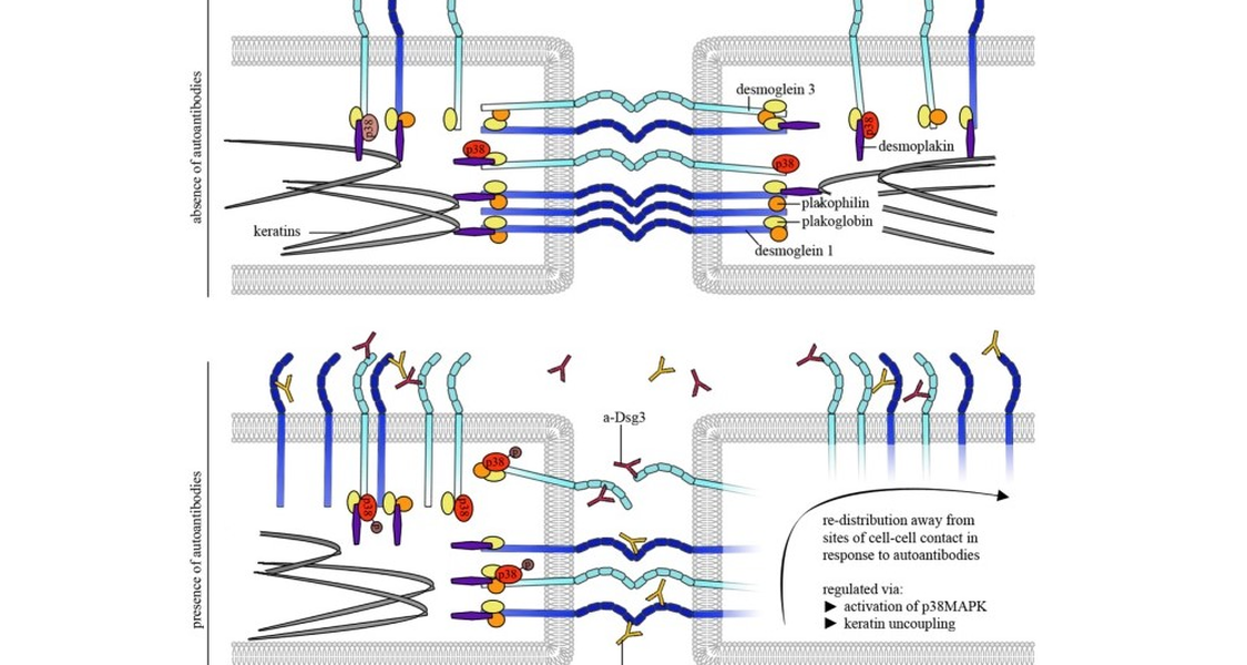
Pemphigus is a bullous autoimmune disease in which autoantibodies directed against the desmosomal cadherin desmoglein (Dsg) 1 and 3 lead to loss of intercellular adhesion in the epidermis and mucous membranes. Dsgs are transmembrane proteins which maintain proper adhesive function of desmosomes by forming Ca2+-dependent homo- and heterophilic interactions with their extracellular domains. Mechanisms contributing to loss of intercellular adhesion in pemphigus comprise direct inhibition of Dsg3 interactions as well as dysregulation of various signalling pathways upon autoantibody binding. Further, binding properties of Dsg1 and 3, both known to act homo- and heterophilically, were changed upon autoantibody binding although the mechanisms leading to these alterations are not yet elucidated. Thus, we aim to characterize the interactions Dsg1 or 3 undergo and their importance for pemphigus pathogenesis using atomic force microscopy.
In the current project we will investigate which heterophilic interactions Dsg1 and 3 molecules undergo with other desmosomal cadherins as well as with the classical cadherin E-Cadherin. We will determine interaction mechanisms of homo- and heterophilic Dsg interactions using mutants of Dsg3 extracellular domain to show their impact on binding properties of the molecule and its distribution and clustering in keratinocytes. Further, we will investigate the effect of pathogenic aDsg1 and aDsg3 antibodies on heterophilic Dsg interactions and characterize upregulation and altered distribution of heterophilic interaction partners of Dsg1 and 3 upon pemphigus autoantibody treatment to elucidate their role in possible compensatory mechanisms in pemphigus.
Next, we will study the role of desmosomal hyper-adhesion on loss of intercellular adhesion and in pemphigus wound healing. Desmosomal hyper-adhesion represents a strong adhesive state in which desmosomal cadherins become independent from extracellular Ca2+. Hyper-adhesion was shown to be important for both, pemphigus pathogenesis and wound healing. Thus, we aim to elucidate the mechanisms underlying this phenomenon. To do so, we will apply the ex-vivo skin model to show which Dsg isoforms acquire a hyper-adhesive state. Further, by using atomic force microscopy we will describe the isoform-dependent correlates of desmosomal hyper-adhesion on the level of single molecule interaction. Furthermore, we will study the role of hyper-adhesion in autoantibody-induced loss of intercellular adhesion and compromised wound healing in pemphigus.
Taken together, in the current project we aim to elucidate the impact of heterophilic interactions as compensatory mechanisms in pemphigus. In addition, we will characterize the single molecule correlate of desmosomal hyper-adhesion. We are convinced that both approaches contribute to a more concise knowledge on regulation of desmosomal adhesion and mechanisms leading to loss of intercellular adhesion in pemphigus.
Contact
Dr. Franziska Vielmuth
Project leader TP6new
Department of Anatomy
Ludwig Maximilians University Munich
Pettenkoferstr. 11
D-80336 Munich
Tel +49 89 2180 72700
Fax +49 89 2180 72602
Email: Franziska.Vielmuth@med.uni-muenchen.de