Hauptinhalt
Ausstattung
Konfokale Laser Scanning Mikroskope:
Leica STELLARIS 8 FALCON (Biologiegebäude, Karl-von-Frisch-Str. 8)
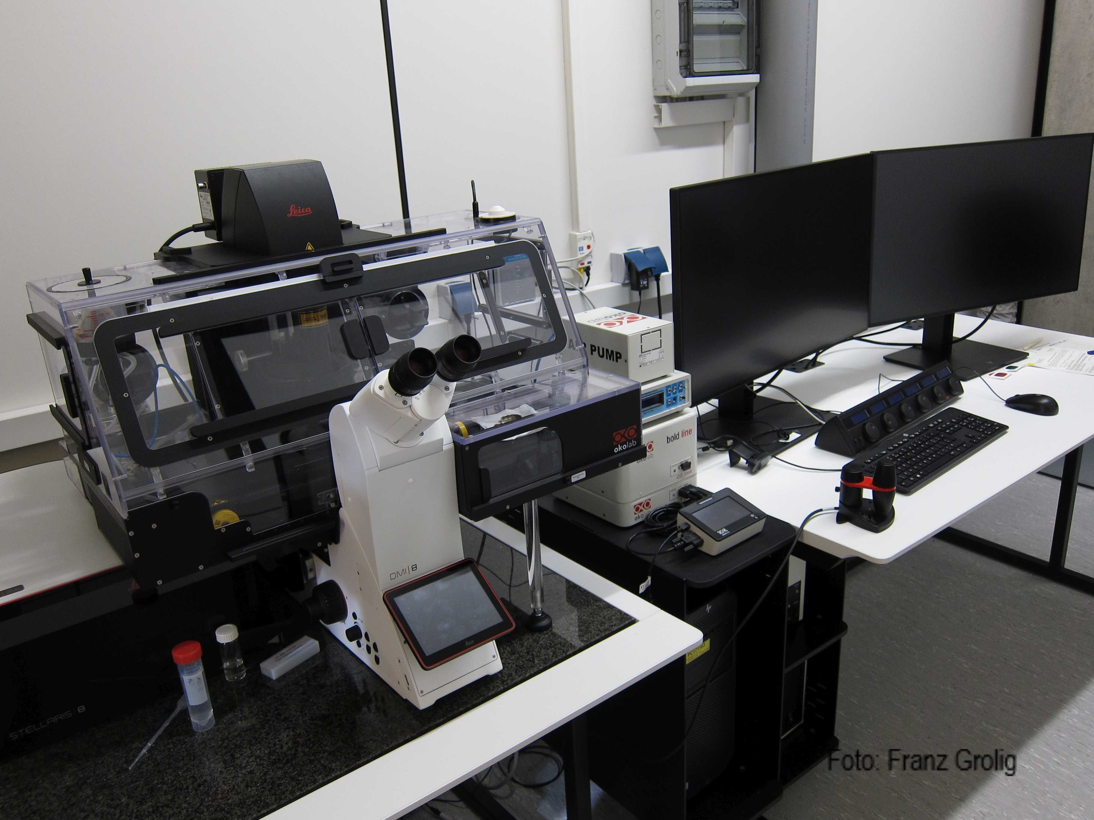
· DMi8 CS inverses Mikroskop mit Super Z Galvo Tisch and Okolab Inkubationsbox
· Diodenlaser 405 nm, 488 nm; Weißlichtlaser 440 – 790 nm
· Detektoren: 2 x HyD S, 2 x HyD R, 1 x HyD R (Detektionsbereich 440 – 850 nm)
· FOV Scanner
STELLARIS 8 Objektive:
· PL APO 20x/0,75 IMM CORR
· HC FLUOTAR L 25x/0,95 X VISIR 0,17
· PL APO 40x/1,10 W mot CORR
· PL APO 63x/1,30 GLYC CORR DIC
STELLARIS 8 Anwendungen:
· Langzeitbeobachtung lebender Zellen bei kontrollierter Temperatur und CO2 Konzentration
· FRAP
· FRET
· FLIM Lösungen: TauSense (qualitativ) und FALCON (quantitativ)
· Photonenzählung (HyD X und HyD R)
· Navigator
· LIGHTNING (Deconvolution)
· DSE (Dynamic Signal Enhancement)
· Lambda Scans
· Live Data Mode
Leica TCS SP5 (Biologiegebäude, Karl-von-Frisch-Str. 8)
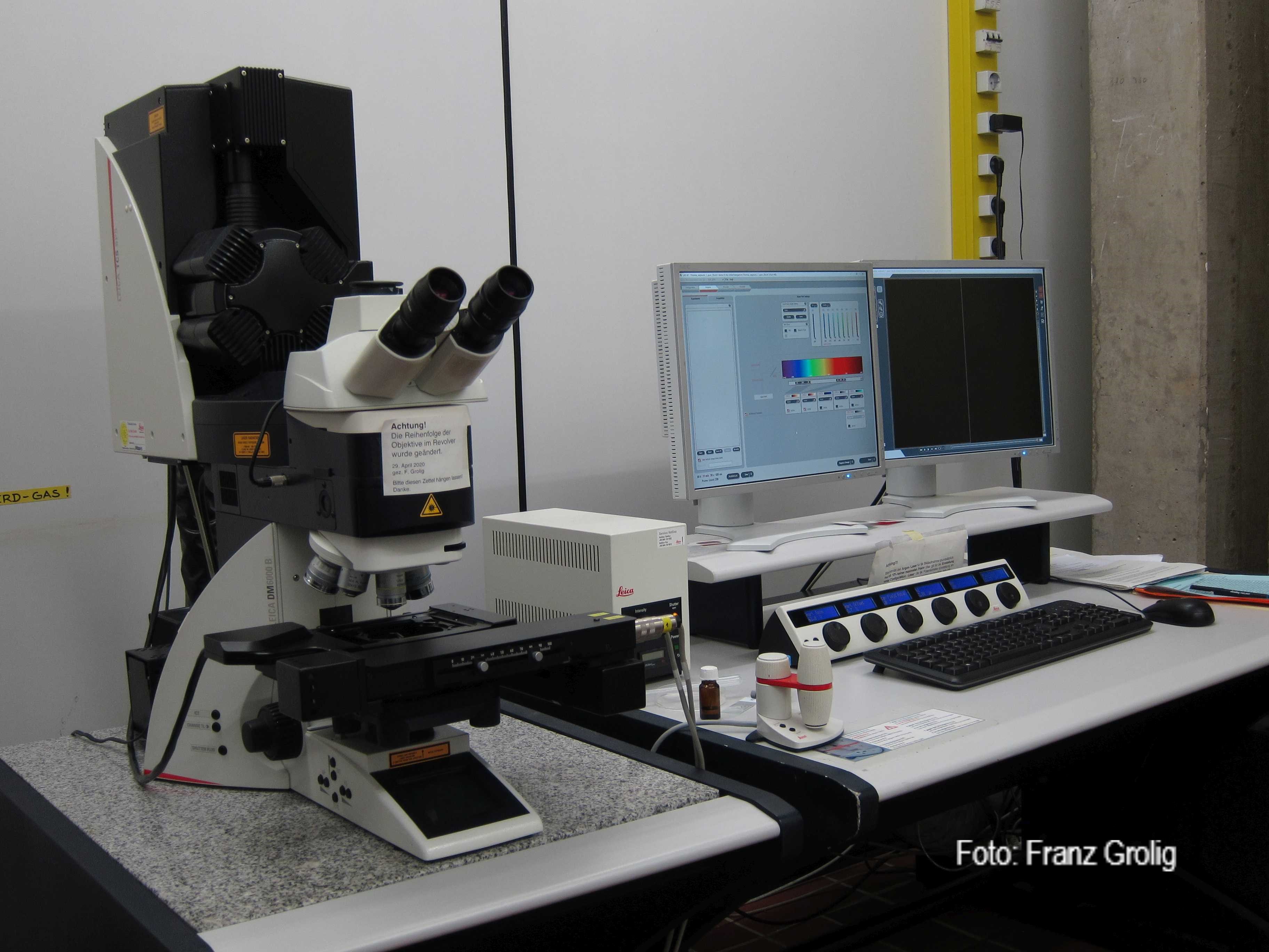
· DM6000 B aufrechtes Mikroskop mit Z Galvo Tisch
· Laser: UV Diode 405 nm; Ar 458, 476, 488, 496, 514 nm; DPSS 561 nm, HeNe 594 nm, HeNe 633 nm
· Detektoren: 5 x PMT (Detektionsbereich 400 – 800 nm)
· FOV und Resonanz Scanner
SP5 Objektive:
HC PL FLUOTAR 10x/0,30
· HC PL APO 10x/0,40 IMM CS
· HCX PL APO 20x/0,70 lmm Corr Lbd. bl. HCX
· PL APO 40x/1,25-0,75 Oil Lbd. bl. HCX PL
· APO 63x/1,40 oil PH3
· HCX PL APO 63x/1,3 GLYC CORR CS 21
SP5 Anwendungen:
· Multidimensionale Aufnahme und Analyse von Bildserien
· FRAP
· FRET
· Mark & Find
· Lambda Scans
· Live Data Mode
Zeiss LSM980 with extra Rapp UV-Box (MZVG 06C06, Hans-Meerwein-Str.6)
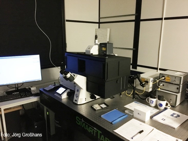
· Axio Observer, inverse, motorized XY, 10 nm focus steps, Z-piezo with 500 µm range, motorized filter ring, condensor with shutter, black incubator box
· Solid state laser 405nm, 488nm, 514nm, 561nm, 594nm, 639nm
· Airyscan 2 array detector, Spectral GaAsP detector, 2x photomultiplier detector
· Photoncounting mode
· Autofocus, focus control in longterm imaging
· Second independent scan module (Rapp UGA-42 caliburn)for illumination via epiport
· UV laser box: 355/42 nm (pulsed), 375/70 nm (continuous wave)
· large system table NLO table, 1.5x1.8m
· second computer, linked to system for real-time data transfer and processing
LSM980 objectives:
· C-Apochromat 40x/1.2 W
· Plan-Apochromat 63x/1.4 oil
· LCI Plan-Apochromat 63x/1.2 multi immersion
LSM980 special applications
o Recording based highresolution imaging down to 120 nm lateral
o Multiplex imaging (up to eight lines simultaneously)
o FRAP
o fluctuation analysis and FCS
o Laser ablation with pulsed UV laser, junctions cuts, wounding, organelle ablation, microsurgery
o UV uncaging with 375 nm UV laser, e.g. NP-EGTA für Ca2+ uncaging
o realtime processing and data transfer to second computer
o longterm (multiple hours) time lapse imaging with readjusted axial focus
o tiling of large objects (mm scale)
Epifluoreszenz Mikroskope:
Zeiss AxioObserver Z1 (Biologiegebäude, Karl-von-Frisch-Str. 8)
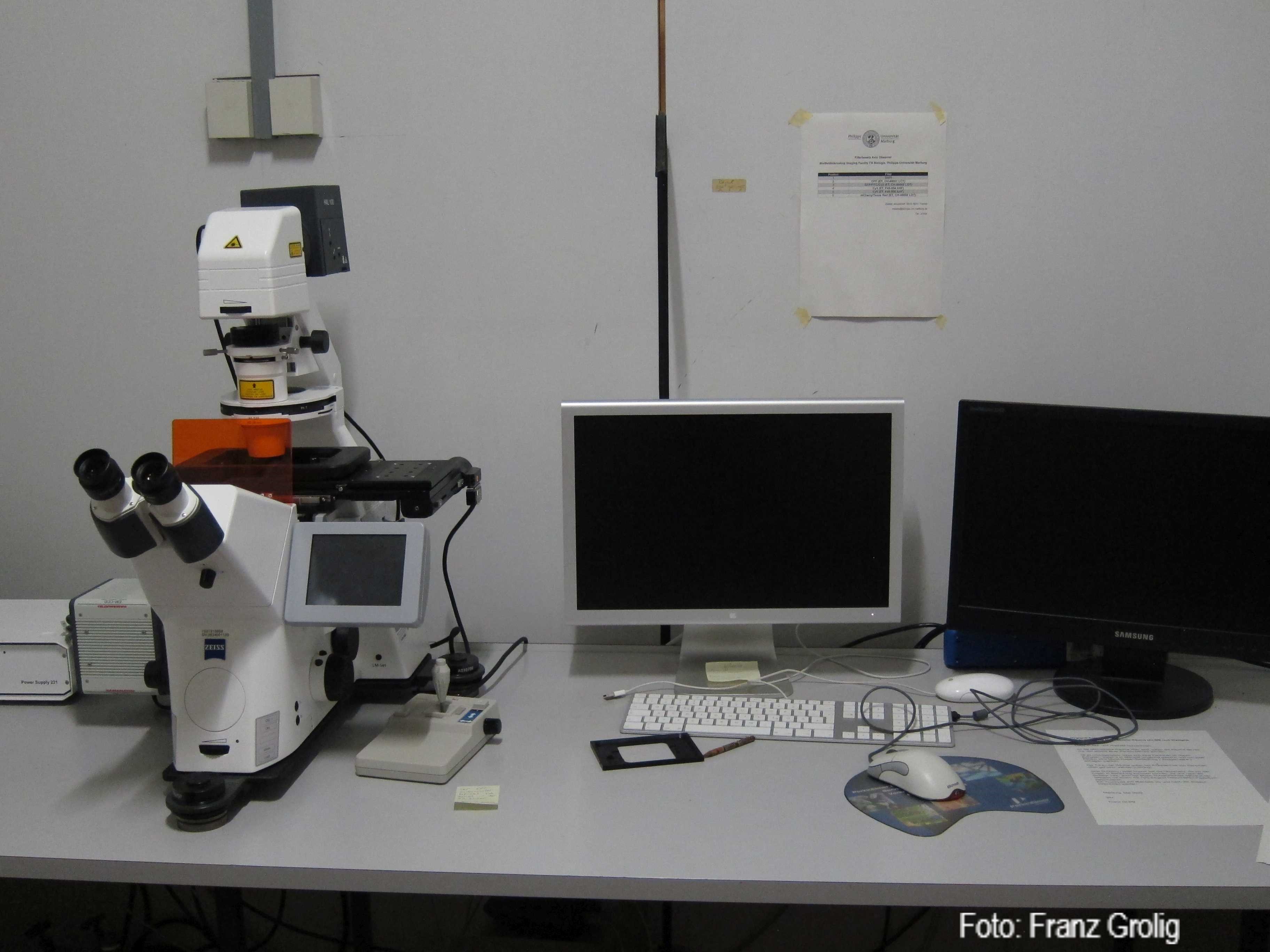
· inverses Mikroskop mit motorisiertem xyz Tisch
· Filter: DAPI, GFP, YFP, CY3, CY5
· Kamera: Zeiss Axiocam 305 mono R2
AxioObserver Z1 Objektive:
· Plan Neofluar 5x
· Plan Neofluar 10x
· Plan Neofluar 20x
· Plan Neofluar 40x (Öl)
· Plan APO 63x
AxioObserver Z1 Anwendungen:
· Echtzeitbeobachtung fluoreszierender Moleküle
· ratio imaging
· FRAP
· FRET
Zeiss AxioObserver (MZVG 06C06, Hans-Meerwein-Str.6)
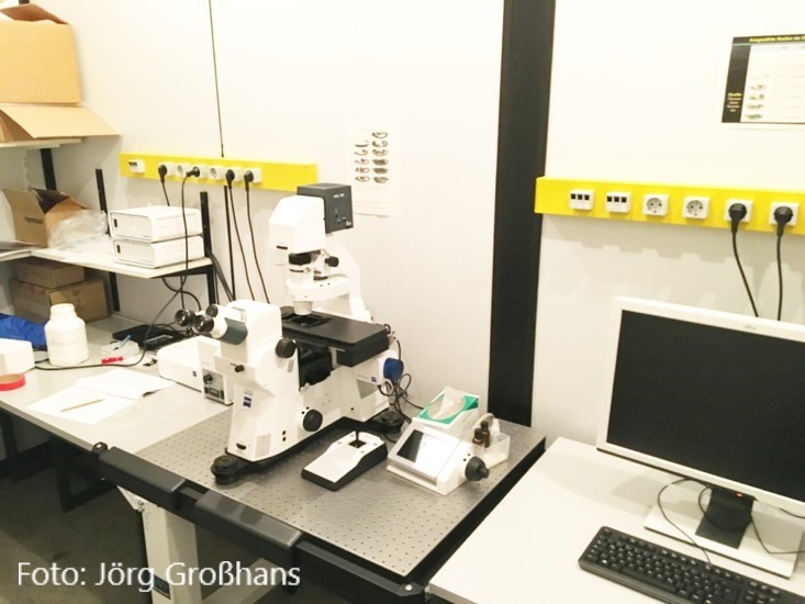
· AxioObserver Z1, inverted, differential interference contrast, phase contrast, motorized stage with joystick
· Detectors: AxiocamMR, emCCD (photometrics evolve 512), ApoTome
AxioObserver objectives:
· 10x
· 20x
· 40x
· 63x/oil
· 100x/oil
AxioObserver special applications:
o Optical sectioning with ApoTome
o simultaneous time lapse recordings (DIC, wide field) with tilling and position module
Zeiss AxioPlan (MZVG 06C06, Hans-Meerwein-Str.6)
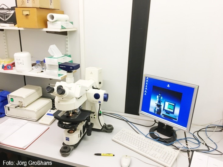
· Axioplan 2, upright, differential interference contrast, phase contrast, darkfield optics
· Detectors AxiocamMR, ApoTomeI
Axioplan objectives:
· 10x
· 20x
· 40x/oil
· 63x/oil
· 63x/PH/oil
· 100x/oil
Axioplan special applications:
o Optical sectioning with ApoTome
Gefördert durch:

