Main Content
Figure 3
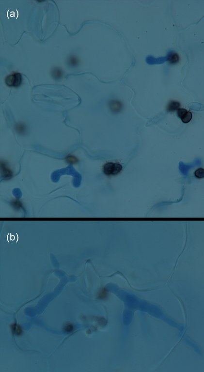
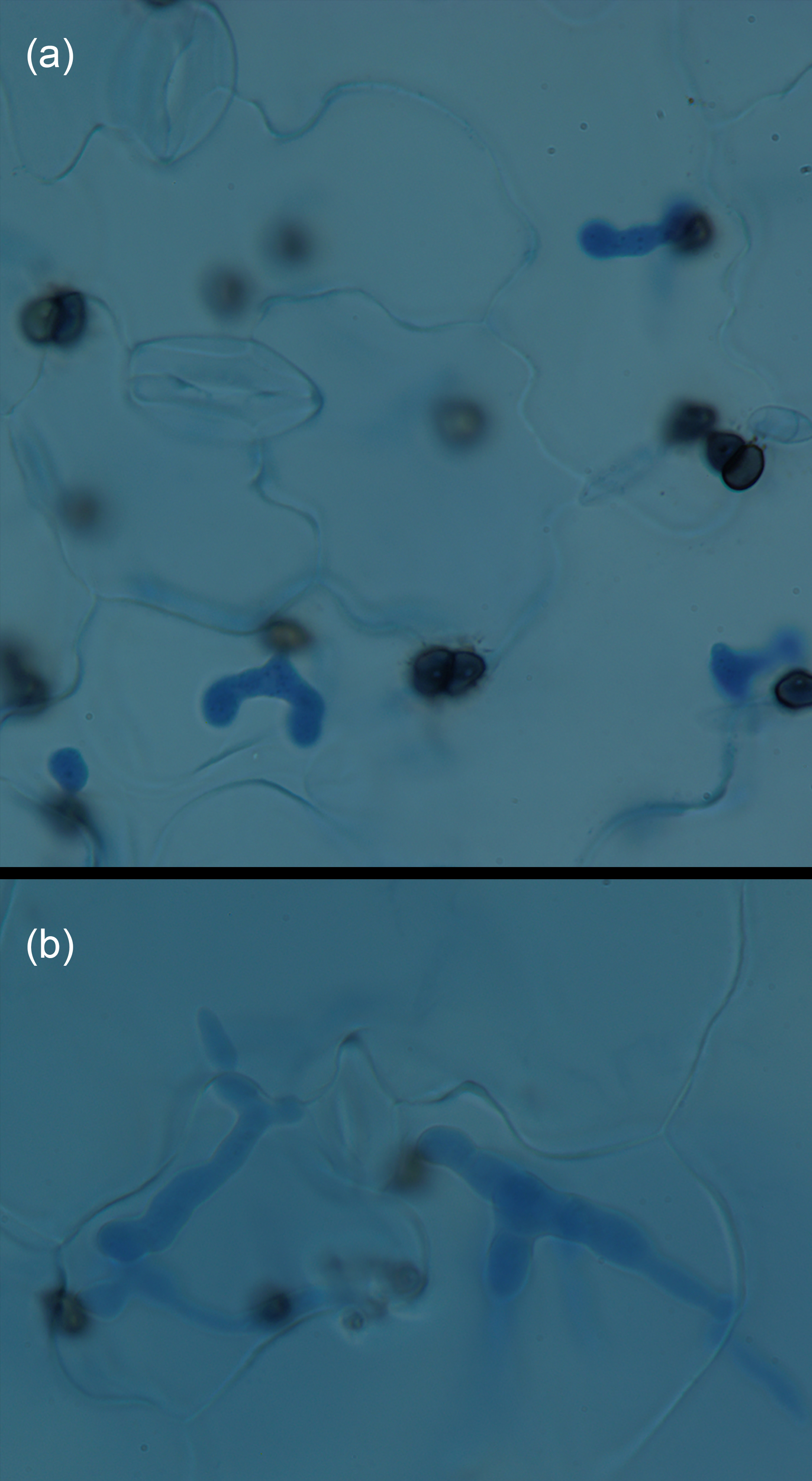
Figure 3. Episodes of the Colletotrichum higginsianum life cycle on infected Arabidopsis leaves.
Top (a): Early infection process (2 days post inoculation) - melanised brownish appressoria (penetration organs on the leaf surface) and lobed, blue stained biotrophic hyphae (inside the penetrated epidermis cells) are abundant.
Bottom (b): Intermediate infection process (3 days post inoculation) - melanised brownish appressoria are out of focus, biotrophic hyphae have expanded to the cell margins of the initially penetrated cell, while necrotrophic secondary hyphae are emerging into the adjacent epidermis cells
(click to enlarge figure)