Main Content
3D Morphology of Random Porous Media
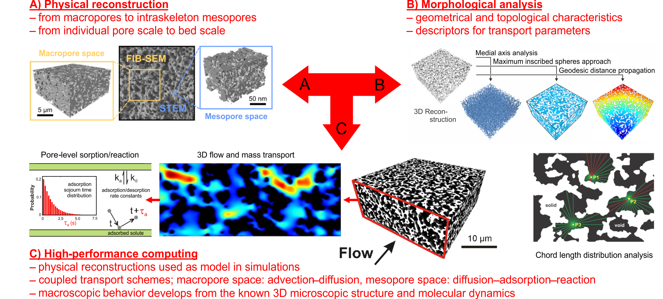
Materials with a hierarchically structured pore space are employed as support structures in processes that require a large surface area for physical or chemical interactions combined with efficient molecular transport to and from the active sites, which applies to mass storage, energy applications, catalysis, and chemical separations. A network of larger macropores (with µm to mm pore size) allows percolation of a mobile phase (gas, liquid, or supercritical fluid) under an external force (gradients in electrical potential or pressure) applied to the material, enabling advective transport of the bulk fluid and solute molecules. A second network of smaller pores (meso- and/or micropores), accessible to solvent and solute molecules only by diffusion, provides a large surface area covered with functionalities tailored to a certain application. Thus, the design of improved morphologies exposing functional interfaces is a major challenge in the advanced engineering of processes relying on selective adsorption or reaction, and the preparation of materials with a hierarchically structured pore space in particular is fundamental to the operation of flow-driven processes for separation, storage, and catalysis.
Porous media have numerous applications, e.g., in battery electrodes, heterogeneous catalysis, and chromatographic separations. Their performance is strongly related to their morphology since efficient mass transfer is crucial to avoid transport limitations. An accurate characterization of porous materials enables to investigate morphology–transport relationships and, with this, to rationally design optimized structures to address the required transport properties of the application. Physical reconstruction emerged as ideal technique for this morphological characterization using both statistical methods as well as advanced mass transport and 3D flow simulations on a high-performance supercomputing platform to identify the performance-limiting morphological properties. As imaging techniques, we use focused ion beam scanning electron microscopy (FIB-SEM) and confocal laser scanning microscopy (CLSM) for macropore space and scanning transmission electron microscopy (STEM) for mesopore space.
Highlighted publications:
- S. Bruns, U. Tallarek
Physical reconstruction of packed beds and their morphological analysis: Core–shell packings as an example.
Journal of Chromatography A 2011, 1218, 1849–1860. DOI: 10.1016/j.chroma.2011.02.013
- K. Hormann, V. Baranau, D. Hlushkou, A. Höltzel, U. Tallarek
Topological analysis of non-granular, disordered porous media: Determination of pore connectivity, pore coordination, and geometric tortuosity in physically reconstructed silica monoliths.
New Journal of Chemistry 2016, 40, 4187–4199. DOI: 10.1039/C5NJ02814K
- T. Müllner, K.K. Unger, U. Tallarek
Characterization of microscopic disorder in reconstructed porous materials and assessment of mass transport-relevant structural descriptors.
New Journal of Chemistry 2016, 40, 3993–4015. DOI: 10.1039/C5NJ03346B